Anatomy Of Upper Leg Muscles And Tendons : Anatomy Of Knee - It consists of a posterior, anterior and lateral compartment.
byAdmin•
0
Anatomy Of Upper Leg Muscles And Tendons : Anatomy Of Knee - It consists of a posterior, anterior and lateral compartment.. Muscles, either individually or in groups, are supported by fascia. On the anterior side, the most prominent of the muscles are the sartorius muscle and the four muscles that make up quadriceps muscle group (the quads.) More specifically, this beautifully illustrated anatomy chart includes neck and shoulders, multiple views of the back and spine, and frontal views of each muscular extremity of the human body. Weak adductor muscles may cause knee instability and adductor strain (2). The muscles in the posterior compartment of the thigh are collectively known as the hamstrings.
Weak adductor muscles may cause knee instability and adductor strain (2). When a muscle contracts, the tendon pulls on the bone causing the joint to move. Rectus femoris these four muscles come together to form a single tendon, which inserts into the patella, or kneecap. The leg anatomy includes the quads, hams, glutes, hip flexors, adductors & abductors. Possibly the most important tendon in terms of mobility is the achilles tendon.
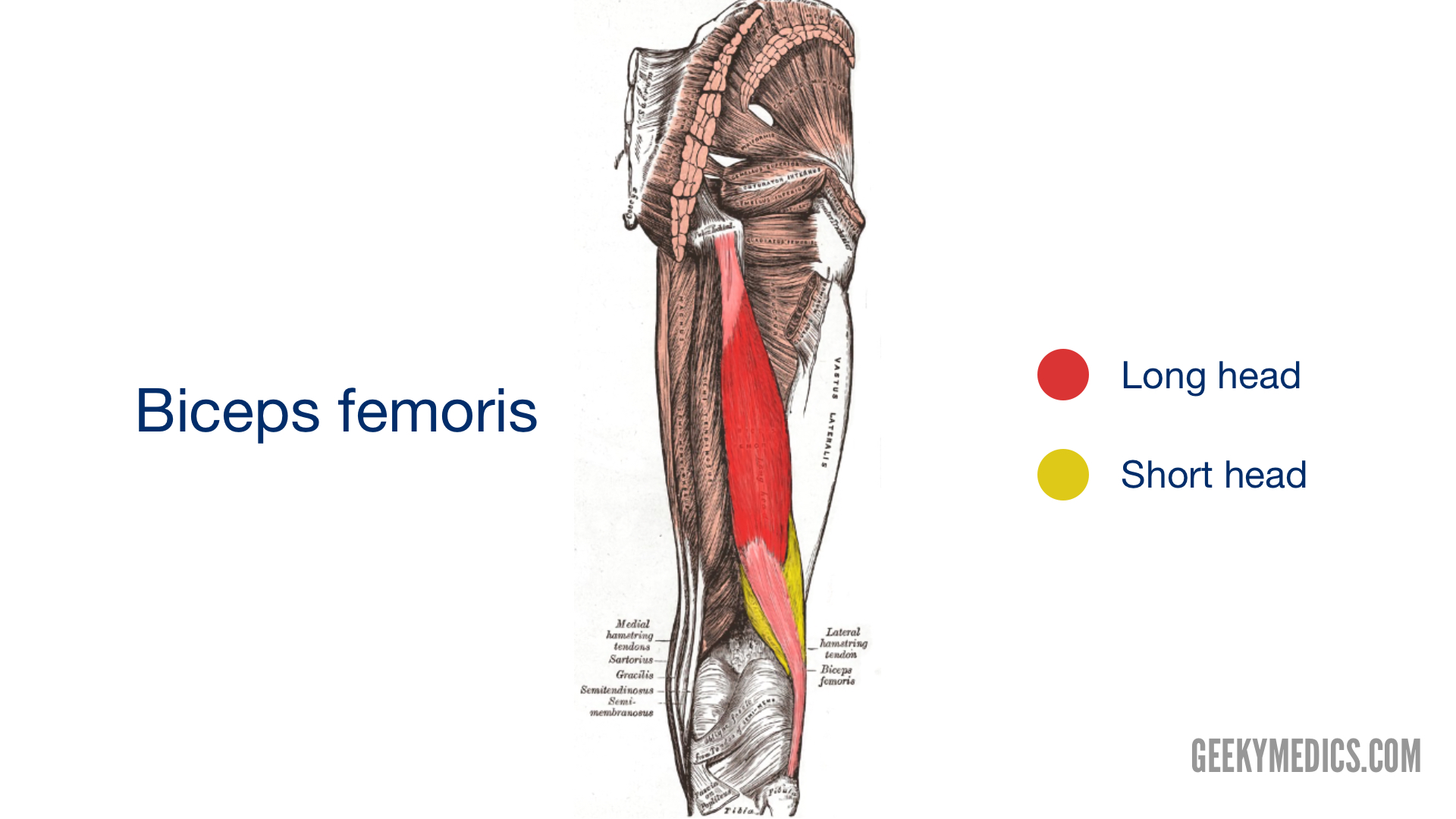
Muscles Of The Posterior Thigh Hamstrings Geeky Medics from geekymedics.com Thigh muscles are responsible for allowing normal gait and proper lower extremity function (1). Possible ruptures of ligaments, muscles and tendons. The thigh bone or femur and the pelvis which is made up of three bones called ilium, ischium, and pubis. This is the group of muscles that you often see body builders flexing, which protrude just above the knee and take up most of the upper leg. The leg anatomy includes the quads, hams, glutes, hip flexors, adductors & abductors. Tendons attach the muscles to each other. Like the forearm, the upper leg, or thigh, has a dense arrangement of many muscles. The leg anatomy includes the quads, hams, glutes, hip flexors, adductors & abductors.
The knee joint is most significantly affected by two major muscle groups:
The largest tendon in the knee is the patellar tendon. Tendons attach the muscles to each other. When the muscle contracts, the tendons are pulled, and the bone is moved. Anatomy_of_leg_muscles_and_tendons 2/7 anatomy of leg muscles and tendons mobi anatomy of leg muscles and tendons anatomy of leg muscles and leg muscles (musculi cruris) anatomically, the leg is defined as the region of the lower limb below the knee. Rectus femoris muscle, one of the. They're found on the ends of muscles, where they help attach muscle to bone. The medial, or towards the middle of the body, upper leg. Other muscles of the anterior (front) thigh include the pectineus, sartorius,. The thigh bone or femur and the pelvis which is made up of three bones called ilium, ischium, and pubis. Muscles, either individually or in groups, are supported by fascia. Tendons also help to provide stability around the foot and ankle. It is comprised of two bones: More specifically, this beautifully illustrated anatomy chart includes neck and shoulders, multiple views of the back and spine, and frontal views of each muscular extremity of the human body.
The largest tendon in the knee is the patellar tendon. Muscle anatomy cross section 12 photos of the muscle anatomy cross section anatomical cross section of muscle, calf muscle anatomy cross section, hamstring muscle anatomy cross section, muscle cross section strength, thigh muscle anatomy cross sectional, human muscles, anatomical cross section of muscle, calf. As group, these muscles act to extend at the hip, and flex at the knee. Ligaments are soft tissue structures that connect bones to bones.a joint capsule is a watertight sac that surrounds a joint.in the hip, the joint capsule is formed by a group of three strong ligaments that connect the femoral head to the acetabulum. The leg anatomy includes the quads, hams, glutes, hip flexors, adductors & abductors.
Upper Leg And Lower Leg Muscle Anatomy from www.anatomynote.com Iliopsoas muscle, a hip flexor muscle that attaches to the upper thigh bone. Ligaments, tendons, and muscles play an important role in the function of the hip. It is comprised of two bones: Nerves and blood vessels that supply the bones and muscles of the hip. It runs from your inner thigh to the quad tendon. These three muscles attach to the achilles tendon, and they all aid with plantarflexion. The anterior, or front upper leg muscles are the quadriceps. They are attached to the femur (thighbone), tibia (shinbone), and fibula (calf.
The gastrocnemius muscle supersedes its function.
This area consists of bones, muscles, tendons, and nerves that all work together to allow the leg to function. Tendons attach the muscles to each other. They are attached to the femur (thighbone), tibia (shinbone), and fibula (calf. Weak adductor muscles may cause knee instability and adductor strain (2). When a muscle contracts, the tendon pulls on the bone causing the joint to move. Tendons are also bands of connective tissue. It consists of a posterior, anterior and lateral compartment. Large ligaments, tendons, and muscles around the hip joint hold the bones (ball and socket) in place and keep it from dislocating. Ligaments, tendons, and muscles play an important role in the function of the hip. There are a number of tendons located in the foot and ankle all responsible for different ankle, foot and toe movements. They consist of the rectus femoris, vastus intermedius, vastus lateralis and the vastus medialis. Muscles, either individually or in groups, are supported by fascia. The anterior, or front upper leg muscles are the quadriceps.
Like the forearm, the upper leg, or thigh, has a dense arrangement of many muscles. Tendons are also bands of connective tissue. It consists of a posterior, anterior and lateral compartment. These three muscles attach to the achilles tendon, and they all aid with plantarflexion. Upper two thirds of the medial margin and proximal margin of the patella, medial condyle of the tibia, and investing deep fascia of the leg with the tendons of vastus intermedius, lateralis, and rectus, and through the patellar ligament onto the front of the tibial tuberosity.
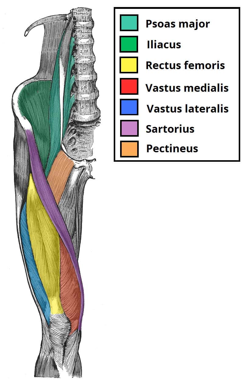
Muscles Of The Anterior Thigh Quadriceps Teachmeanatomy from teachmeanatomy.info The posterior, or back, leg muscles for the upper leg are the hamstrings. Proximal margin and deep surface of. From its origin, the vastus lateralis courses down your lateral thigh and inserts as part of the lateral quadriceps tendon on the tibal tubercle. Related posts of muscles and tendons of the leg muscle anatomy cross section. Muscle anatomy cross section 12 photos of the muscle anatomy cross section anatomical cross section of muscle, calf muscle anatomy cross section, hamstring muscle anatomy cross section, muscle cross section strength, thigh muscle anatomy cross sectional, human muscles, anatomical cross section of muscle, calf. The quadriceps muscles provide strength and power with knee extension (straightening). Ligaments, tendons, and muscles play an important role in the function of the hip. The knee's anatomy consists of many structures from the bones, tendons, and ligaments to the cartilage and muscles to help the knee function.
When the muscle contracts, the tendons are pulled, and the bone is moved.
Thigh muscles are responsible for allowing normal gait and proper lower extremity function (1). There are a number of tendons located in the foot and ankle all responsible for different ankle, foot and toe movements. Like the forearm, the upper leg, or thigh, has a dense arrangement of many muscles. This is the group of muscles that you often see body builders flexing, which protrude just above the knee and take up most of the upper leg. Muscles, either individually or in groups, are supported by fascia. It consists of a posterior, anterior and lateral compartment. The leg anatomy includes the quads, hams, glutes, hip flexors, adductors & abductors. They're found on the ends of muscles, where they help attach muscle to bone. Related posts of muscles and tendons of the leg muscle anatomy cross section. The gastrocnemius muscle supersedes its function. Ligaments are soft tissue structures that connect bones to bones.a joint capsule is a watertight sac that surrounds a joint.in the hip, the joint capsule is formed by a group of three strong ligaments that connect the femoral head to the acetabulum. The muscle fibers descend through the anterior compartment of the leg and converge to a tendon before the ankle. The knee's anatomy consists of many structures from the bones, tendons, and ligaments to the cartilage and muscles to help the knee function.
Tendons attach the muscles to each other upper leg muscles and tendons. Upper leg tendon anatomy muscles of the anterior thigh quadriceps teachmeanatomy the peroneal tendons are in the feet and provide balance and stability during movement leroy hunley from i0.wp.com however, hamstring pulls can also occur at any place along the hamstring muscle bellies or in the tendons that attach the muscles to the bones.